Preceeding pre-registration, a pilot was conducted to finetune the original adaptation of the SART for the purposes of this research:
Method
Piloting for the experiment was carried out on 4 younger participants (2 F). They carried out a preliminary version of the SART task, consisting of 160 go and 40 nogo trials, 13 minute length in total. Most participants also had baseline neural signal recorded and eyes closed recorded.
Analysis
The data was epoched to -1500ms – 1500ms, resampled to 1000Hz in one case and filtered 1-40Hz with eye blinks removed as the first component of an ICA. Observations were made based on visual inspection of data which was run through basic fieldtrip analysis.
Results
Behaviourally, participant behaviour appeared similar to the online pilot done previously - Nogo accuracy: 81.25% (SD = 16.52%), go accuracy: 99.84% (SD = 0.3%). However, reaction times were much slower to the point that of raising suspicion of technical error: 454.27ms (SD = 177.44ms). Finally, there was significant anticipation error, defined as responses before 150ms of stimulus onset: 15.5% (SD = 19.40%). Consequently, the pilot showed that adjusting trial length impacted reaction times.
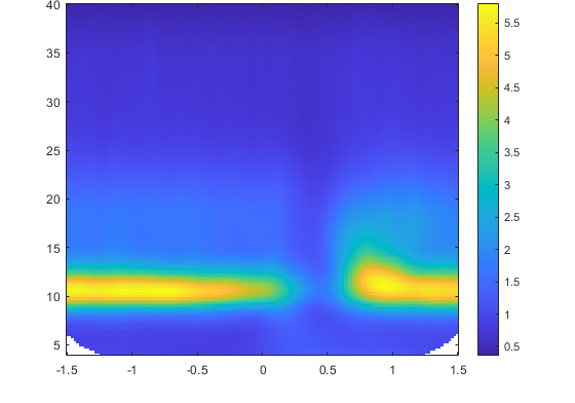
Before Baseline, there appeared to be a significant dip in alpha wave oscillation in the segment 200 – 600ms, see Fig. 1. The segment -700ms to -200ms was used for Baseline.
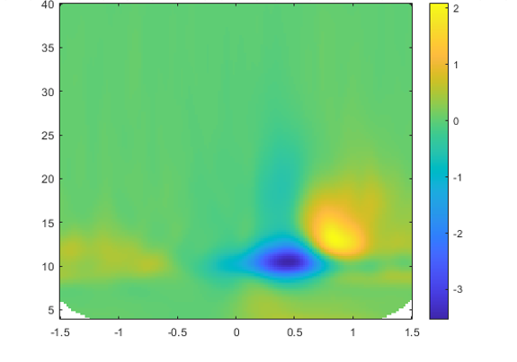
 After Baseline, there was a decrease in alpha oscillation in the 200 – 600ms followed by an increase in lower beta oscillation in the frame 600 – 1000ms. There was some indication of early theta oscillation in the segment (200 – 600ms), see patterns in Fig. 2.
 It seemed that the alpha decrease was primarily parietal (Pz – P6, POz – PO8) whereas the beta increase was more frontal (F1 – F4, AFz – AF4). There was also a small increase in theta both frontally and parietally, in line with latest research (McMackin, 2021).
Overall, the predominant signal was the decrease in alpha level, by several orders of magnitude (10x).
The relative increases in beta were stronger in go trials, otherwise it inference was difficult about the difference in alpha. It is possible that go trial decrease in alpha was more prevalent, especially in the latest segment (1000ms – 1400ms).
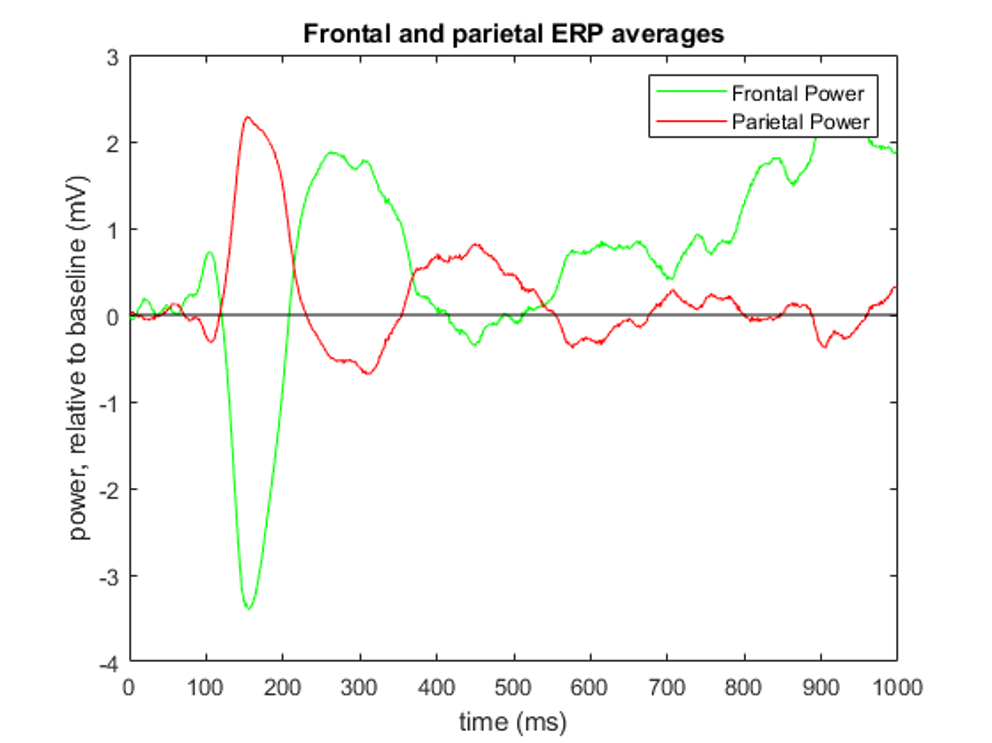
ERP analysis was difficult and inconclusive beyond early visual peaks. Also, the parietal and frontal areas greatly contrasted, see Fig. 3.
 The recess in alpha activity was concluded as the single most important feature and should ideally be perfectly localised in the subsequent study, perhaps choosing to employ fewer candidates. There was also a beta rebound, stronger in the go trials, corresponding to motor action.
The ERPs were not addressed in great detail, but showed indication of active visual processing in the parietal scalp locations.